Scientists map basal ganglia in most detailed atlas yet
An international team profiled 25.5 million brain cells to create the most detailed atlas of the basal ganglia, published on 12 November.
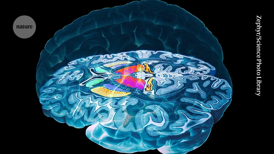 Photo: Nature
Key points
Researchers published the most detailed atlas of the basal ganglia, profiling 25.5 million cells and identifying new cell types linked to schizophrenia and Parkinson's disease.
An international consortium of more than 200 scientists published the most detailed atlas yet of the basal ganglia, a brain region tied to Parkinson's disease, schizophrenia and addiction. The work, released on 12 November, profiles 25.5 million brain cells from humans, macaques and marmosets. The atlas is described in a collection of 26 papers published in Cell, Cell Reports, Cell Reports Methods, Cell Systems, Cell Genomics and Neuron.
The basal ganglia are groups of cells that sit deep in the brain and process motor, cognitive, mood and decision-making information. They are linked to neurodegenerative and psychiatric conditions from Parkinson's and Huntington's disease to schizophrenia, bipolar disorder and addiction. The new atlas is part of the BRAIN Initiative Cell Atlas Network, a US$500-million project launched in 2022 by the US National Institutes of Health.
A cross-species reference
The atlas contains 21 million cells from the brains of six humans, three million cells from three rhesus and cynomolgus macaques, and 1.5 million cells from two marmosets. The researchers also used data from previously published maps of the whole mouse brain. By comparing the basal ganglia between the primate species, they identified 61 cell types that have been conserved over 76 million years.
One study examined 1.1 million cells in the striatum, the basal ganglia's largest structure, from 19 deceased human donors. Unlike many other brain areas, the striatum has no neat anatomical boundaries. The researchers identified six zones in the human and macaque striatum with distinct molecular signatures, each containing distinct populations of neurons and glial cells.
New cell types and disease links
"The cells in these zones all look the same under the microscope, but have very strong transcriptional differences and are strikingly consistent from person to person," said co-author Evan Macosko, a neurobiologist at the Broad Institute of Harvard and MIT. Macosko and his colleagues also found that gene expression patterns that distinguished each zone became increasingly similar with age, particularly in brain regions affected by Huntington's and Parkinson's disease.
Another study analysed nearly four million cells in the striatum and prefrontal cortex from 178 deceased adult donors between the ages of 27 and over 90. The researchers found that the cellular make-up of the two brain regions varied between people, particularly for non-neuronal cells known as glial cells. This underscores the need for brain cell atlases to account for individual differences.
The researchers also identified a previously unknown brain cell type linked to schizophrenia and bipolar disorder. The study highlights the importance of a rare neuronal subtype that expresses the serotonin receptor HTR7 and is associated with an elevated risk of schizophrenia. The findings make it possible to trace the disorder's genetic signals back to specific cells, potentially improving research into the disease.
Age-related changes
Another study describes a new version of an existing gene therapy that reduces the tremors characteristic of Parkinson's disease. In animal models, the approach proved more effective and less invasive than treatments currently used in patients. A study led by researchers at the University of California, San Francisco identified significant genetic changes in specific cell types within the basal ganglia that may play a role in autism.
Scientists at the Broad Institute discovered widespread age-related changes in gene expression that affect different cell types in distinct ways. Their findings make it possible to predict a person's age from gene-expression patterns across multiple types of brain cells. For example, the abundance of oligodendrocyte progenitor cells declines by about 40% between the ages of 30 and 80.
The atlas is the latest achievement of the BRAIN Initiative, a major US research program launched in 2014 that has so far received roughly $750 million in funding. Its ultimate goal is to produce a comprehensive catalogue of every cell type in the human brain. "To understand what is truly happening in the brain, we need an accurate list of cell types and to study gene expression in each of them," explains Hongkui Zeng, executive vice president and director of neuroscience at the Allen Institute.
The plan is to follow this map with a similarly detailed version for the entire human brain, a scientific feat that has not yet been accomplished. Ed Lein, executive vice president and director of brain health at the Allen Institute, likened the idea to that of the Human Genome Project. "The ambition is to create some combination between the Human Genome Project and Google Earth, for the brain," Lein said.
Frequently asked questions
What is the basal ganglia?
The basal ganglia are groups of cells deep in the brain that process motor, cognitive, mood and decision-making information. They are linked to conditions such as Parkinson's disease, schizophrenia and addiction.
How many cells were profiled in the new atlas?
The atlas profiles 25.5 million brain cells: 21 million from humans, three million from macaques, and 1.5 million from marmosets. It also uses data from previously published maps of the whole mouse brain.
What new findings were reported?
Researchers identified a new cell type linked to schizophrenia and bipolar disorder, found six molecular zones in the striatum, and showed that gene expression patterns change with age. They also found that cellular composition varies between individuals.